
"High density shadow", "new lymph nodes", "right middle lobe bullae", "liver parenchymal space occupying lesions"
The moment I got the review report, I looked at the medical terms in the report, which made many search friends very panic.
In order to avoid misunderstanding of these medical terms, Huzhu Jun will talk with you today about what the medical terms that look very scary on the breast cancer reexamination report mean and whether they mean that the disease is progressing. Help find friends better understand their condition and what to do next.
1 high density shadow
High density image is a commonly used term in image-assisted examination (such as mammography, CT, etc.).
Most breast cancer has high density or equal density, and a few have low density [1]. But high-density shadow is not necessarily a tumor. It also depends on the surrounding conditions of the high-density shadow, such as whether there is a mass, calcified morphology, structural distortion, asymmetry, intramammary lymph nodes, skin lesions, unilateral dilated ducts and accompanying signs.
In addition, it is necessary to combine the site and other examinations with the age at which the disease is prone and the clinical history. After the specific analysis of the doctor, the targeted examination can get the correct diagnostic results.
2 New lymph nodes
There are many lymph nodes in the lower cervical triangle (neck, jaw, clavicular fossa), armpit, groin, etc. Among them, axillary sentinel lymph nodes are one of the earliest metastatic sites of breast cancer.
The number of lymph node metastases is a very important indicator, which determines the stage of disease, treatment and prognosis. Generally speaking, the more lymph node metastasis, the more serious the disease, the worse the prognosis [2].
Therefore, if multiple new lymph nodes, especially the axillary part, are found in the breast ultrasound report, further examination and treatment should be carried out as soon as possible in combination with the clinical and patient's physical conditions.
3 ground glass nodules in the apical segment of the upper lobe of the lung
The apical segment of the upper lobe of the lung generally refers to the apical segment of the upper lobe of the right lung. Ground glass nodule is an imaging manifestation of chest CT. CT showed that the local areas in the lung tissue had light shadows like ground glass [3].
The detection of ground glass nodules in the lung does not mean that breast cancer must have metastasis, but simply describes the circular or quasi circular lesions found in the process of CT examination.
After the first discovery of ground glass nodules, the general doctor will advise the patients to follow up for 3 months, 6 months and 12 months. During the follow-up, the doctor will compare the results of this follow-up with the previous one to see whether the nodules have increased, whether the density has increased, and whether there are characteristic imaging findings.
In case of enlargement of nodules, increase of solid components or new malignant imaging manifestations during follow-up, it is necessary to shorten the follow-up interval, conduct puncture biopsy to determine the pathological type, and make further intervention [4].
4 right middle lobe bulla
The right lung is divided into upper, middle and lower lobes. Pulmonary bullae, also known as pulmonary bullae, refer to the air bag containing cavities formed in the lungs due to various reasons, which lead to the rupture of pulmonary alveoli with respiratory function due to the increase of intraluminal pressure and the increase of alveolar wall tension to a certain extent, resulting in the fusion of alveoli [5].
The detection of pulmonary bullae does not mean that there is a risk. It is necessary to do a further chest CT or X-ray examination to clarify the number, location, scope, true size of pulmonary bullae and the relationship between the surrounding tissues.
As for the need for treatment. The study points out that asymptomatic pulmonary bullae do not need treatment, patients with chronic bronchitis or emphysema need treatment, and secondary infection can be treated with antibiotics; If the volume of pulmonary bullae is large and accounts for 70% ~ 100% of one side of the thoracic cavity, the patient will have respiratory distress, infection, bleeding and other clinical manifestations, so surgical treatment is required [6].
5 hypoechoic nodule of left supraclavicular fossa
Echo is a commonly used term in breast ultrasound examination, and nodule is not the name of the disease, nor is it used to describe the benign or malignant tumor, but only to describe the small tumor found in the process of breast ultrasound examination.
In breast ultrasound, the common echoes mainly include hyperechoic nodules, hypoechoic nodules, anechoic nodules and weakly echoic nodules. Among them, hypoechoic nodules are the most common and represent the parenchymal nodules of the breast [1]. However, low echo can not confirm that the nodule is malignant or benign. Clinicians need to make a comprehensive analysis in combination with other ultrasonic features and test indicators to determine whether surgery is required.
6 Breast structural disorder
In the process of breast cancer ultrasound examination, molybdenum target examination and nuclear magnetic resonance examination, if the subcutaneous adipose tissue structure of the breast is found to be abnormal, it will be described as breast structural disorder in the report given.
Discover mammary gland structure disorder, did breast cancer relapse?
In fact, in most cases, the main factor leading to breast structural disorder is gland proliferation, and the gland structural disorder caused by local cell malignant transformation accounts for a small proportion. However, if breast structural disorder is accompanied by mass, asymmetric density or calcification, biopsy is required to determine whether it is malignant [1].
7 solid space occupying lesions of liver
The normal liver is divided into two parts: the liver parenchyma and the vascular system (blood vessels and bile ducts). When the abnormal area presents the shape of a nodule or mass, occupying a certain space, resulting in the compression, displacement or invasion of adjacent liver tissue and vessels, it is called liver space occupying [7].
The presence of hepatic space occupying lesions in liver ultrasound examination does not mean that breast cancer has metastases. General doctors will recommend that patients have another liver enhanced magnetic resonance or CT examination to judge its benign and malignant.
In addition, some hematology tests, blood biochemistry and serum tumor markers (serum alpha fetoprotein, serum carcinoembryonic antigen, etc.) may be done to assist in the diagnosis, so as to help judge whether there is the possibility of primary liver cancer or whether breast cancer has liver metastasis.
8 Gallbladder wall thickening and roughness
The normal gallbladder wall is a strong echo line band, smooth, neat and natural, and its thickness generally does not exceed 2 ~ 3mm [8]. If the thickness of the anterior wall of the gallbladder body exceeds 3mm, it can be determined as thickening of the gallbladder wall.
Gallbladder wall thickening is a non-specific pathological manifestation, which is mainly seen in cholecystitis, gallbladder cancer, acute and chronic hepatitis, liver cirrhosis, chronic cardiac insufficiency, chronic renal insufficiency and hypoproteinemia [9].
If the ultrasound of patients with breast cancer shows that the gallbladder wall is thickened and rough, they can be considered as suffering from chronic cholecystitis. It is suggested to find out whether it is caused by primary or secondary diseases. At the same time, if the doctor thinks the condition is serious after evaluation, cholecystectomy should be carried out as soon as possible to avoid affecting the treatment of breast cancer.
Summary
A simple understanding of some professional terms that will inevitably appear in your review report can not only avoid mistaking the relapse of the disease because you do not understand the professional terms, but also have a basic understanding of the disease, and timely consult the clinician when it is really necessary, which is a good time for the best treatment.
Reference material:
[1] Breast cancer Professional Committee of China Anti Cancer Association Guidelines and specifications for the diagnosis and treatment of breast cancer of the China Anti Cancer Association (2021) [j] Chinese Journal of cancer, 2021,31 (10): 954-1040
[2] Li Lin Analysis of related factors of breast cancer metastasis [j] China modern drug application, 2014,8 (14): 79-80
[3] Zhang Haikuan, Jia Ning, Leng ailing Clinical analysis of high-resolution CT, chest plain film and pathology in the diagnosis of pulmonary ground glass nodules [j] Chinese Journal of lung diseases (Electronic Edition), 2020,13 (03): 360-364
[4] Ke Mingyao, chenzhide, Zeng Junli, Yong Yazhi, Lin Liancheng Diagnosis, treatment and ablation of pulmonary ground glass nodules [j] Chinese Journal of thoracic and cardiovascular surgery, 2022,29 (01): 11-22
[5] Jing Siming, duanhailing, zhanghuiqin, huangweiping, Liyanling, Qin Shunyun Clinical significance of ultrasonic examination of gallbladder wall thickening [j] Chinese community physician (medical specialty), 2012,14 (06): 252-253
[6] Hou G,Wang W,Wang QY,et al.Bronchoscopic bullectomy with a one-way endobronchial valve to treat a giant bulla in an emphysematic lung:a case report [J]. Clin Respir J,2016,10 (5):657-660.
[7] Wangqiuping Imaging diagnosis strategy of liver space occupying lesions [j] Chinese Journal of clinicians (Electronic Edition), 2014,8 (01): 1-6
[8] Iwama E,Okamoto I,Yabuuchi H,et al.Characteristics of smoking lung cancer patients with emphysematous bullae [J]. J Thorac Oncol,2016,11(9):1586-1590.
[9] Yang shunshi Clinical significance of B-ultrasound in diagnosing gallbladder wall thickening [j] Chinese Journal of medicine, 2007,31 (4): 313-314
Original link:
http://www.mijian360.com/news/18254.html

Increase knowledge! Help you "decipher" the professional terms in the review report
Abstract:Highdensityshadow,newlymphnodes,rightmiddlelobebullae,liverparenchymalspaceoccupyinglesionsThemomentIgotthereviewreport,Ilookedatthemedicaltermsinthereport,whichmademanysearchfriendsverypanic.
1、All the information in this website is available on the Internet, and all of them contain the original text or the source of quotation. You can check the original text;
2、The information on this website is only for learning and popular science, not for any other purpose. Please read it rationally;
3、If the information contained in this website violates your rights, please contact us, we will delete it at the first time, and thank you for your prompt;;
Course of literature review methods:
1、Open the information of the website, where there is the reference source of the information, and click open;
2、Read or download the literature online, most of which are charged. Please download according to your needs and abilities;
3、Baidu search sci-hub, then you can get unexpected download method;
4、Literature Translation: you can easily read the literature by using the "Zhiyun" literature tool;
5、Online English Literature: you can use Baidu translation and other translation tools, or use "copytranslator" to read online more conveniently;
Famous medical literature databases at home and abroad:
Open The Tool


-
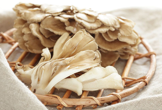
Youyang County, Chongqing City: Maitake under photovoltaic panel
UnderthesunshineofthephotovoltaicAgriculturalIndustrialParkinWanMuvillage,WanMuTown,YouyangCounty,Chongqing,thephotovolt
CloseTo2021-04-07
-
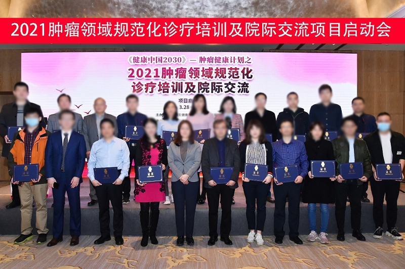
Healthy China 2030
OnMarch28,2021,theprojectHealthChina2030-standardizeddiagnosisandtreatmenttrainingandinterhospitalcommunicationintumorfi
Reports2021-04-01
-
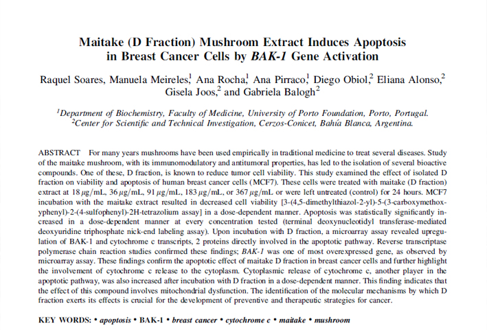
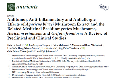
Antitumor, Anti-Inflammatory and Antiallergic Effects of Agaricus blazei Mushroom Extract and the Re
Title:Antitumor,Anti-InflammatoryandAntiallergicEffectsofAgaricusblazeiMushroomExtractandtheRelatedMedicinalBasidiomycet
Research2021-03-31
-
National annual report on cancer surveillance in children
OnMarch20,2021,thefirstmeetingoftheExpertCommitteeonchildrenshematologyandmalignanttumoroftheNationalHealthCommissionand
iHealth2021-03-25
Research / D-Fraction / Experts
- Research Progress of Maitake Active Polysaccharide
-
ResearchProgressofMaitakeActivePolysaccharide
Research2021-01-05
- Experimental study on the effect of Maitake extract on the immune function of mice
-
ExperimentalstudyontheeffectofMaitakeextractonthei
Research2021-01-05
- Experimental study on antitumor effect of Maitake polysaccharide in vivo
-
ExperimentalstudyonantitumoreffectofMaitakepolysac
Research2021-01-04
Maitake News
- 2021 National Cancer Prevention and treatment publicity week
-
TheNationalCancerPreventionandcontrolpublicityweek
Reports2021-04-15
- Healthy China 2030
-
OnMarch28,2021,theprojectHealthChina2030-standardi
Reports2021-04-01
- GB 7096-2014 national food safety standard Edible Fungi and its products

-
GB7096—2014IssuedonDecember24,2014Nationalfoodsaf
Reports2021-03-11
Appliance
Maitake Culture
- Qingyuan Zhejiang is committed to creating the most ecological window of beautiful Zhejiang

-
AsthefirstcountyinChinasecologicalenvironment,tran
CloseTo2020-12-25
- Ancient people's knowledge of Maitake
-
在历史上,我国和日本都属于比较早就认识灰树花的国家,
Culture2020-12-29
- Nutrient composition of Maitake (reference table)
-
日本文部科学省,灰树花营养成分表(参考表)
Culture2021-01-15